Archive for the ‘cystic’ Category

Cystic Intraaxial Supratentorial Brain Lesions
- astrocytoma
- ganglioglioma and ganglioneuroma
- pleomorphic xanthoastrocytoma
- DNET
- sinus mucocele that invaded through cranium
- congenital
Cystic Extraaxial Brain Lesions

- arachnoid cyst
- CSF filled/signal
- middle cranial fossa (MC) > cerebral convexities, basal cisterns, retrocerebellar region
- hypogenesis of adjacent brain
- no enhancement
- low on DWI (like ventricle)
- epidermoid cyst
- has internal matrix, seen in T1
- hyperintense to CSF on FLAIR
- high on DWI (restricted diffusion)

Congenital Cystic Adenomatoid Malformation

- Type 1
- big cysts (2-10 cm)
- Type 2
- multiple small cysts
- renal and cardiac anomalies
- Type 3
- solid
- any lobe
- alveoli replaced by adenomatoid
- surgical resection
- 70% present during 1st week of life
- 10% diagnosed after 1st year
- supplied by pulmonary artery; systemic supply rare
- can involve entire lobe
- prenatal diagnosis
- polyhydramnios
- fetal hydrops
- solid or cystic mass in fetal thorax
- excise affected lobe
Cystic Pediatric Neck Masses
Congenital:
- Thyroglossal duct cyst – base of tongue, hyoid
- 1st Brachial anomalies – between ear and mandible
- 2nd brachial anomalies – mc, type 2 remnant of sinus of His, post to submandibular gland, anterior to scm, AL to vessels, brachiosaurus- otorenal syndrome
- 3rd brachial anomalies – posterior or anterior triangle, cervical thymic cyst extend to mediastinum
- 4th brachial anomalies – pyriform sinus fistula, inflammation next to thyroid
- DermoId cysts – midline, paramidline, orbit, oral, nasal
- Vascular – lymphatovenous malformations, locular, FFlevels
Acquired:
- Suppuratibe adenopathy/abscess
- Necrotic mets
Thin Walled Lung Cysts
- Eosinophilic granuloma
- Lymphangioleiomyomatosis (LAM)
- upper
- uniform size
- women in reproductive years
- can rupture –> pneumothorax
- Tuberous sclerosis
- Neurofibromatosis
- LIP
histiocytosis X
- peribronchiolar distribution
- nodules become cysts
- ~ smoking
- upper lobes
- CPA spared
cystic lung disease
Pericardial Cyst

- fluid filled cardiophrenic angle mass
- R>L
- attached to parietal pericardium; do not communicate with pericardial space
Choledocal cyst

- congenital focal or diffuse dilation of bile ducts
- females
- 5 categories
- type 1 cysts
- 80-90%
- saccular or fusiform
- type 2
- diverticula of duct
- type 3
- choledochoceles
- dilation of terminal, intraduodenal portion of CBD
- type 4
- intrahepatic and extrahepatic cysts
- type 5 = Caroli’s disease
- multiple cystic dilations of the intrahepatic bile ducts
- ~ medullary sponge kidney, AR PCKD
- complications: liver abscess, portal vein thrombosis, biliary cirrhosis with portal hypertension
- 100% risk of cholangiocarcinoma
- MRCP, cholangiography, ERCP shows biliary origins of cysts
- type 1 cysts
- marked biliary stasis: get infection, inflammation, stone disease
- risk of cholangiocarcinoma with age
- in neonates, disorder ~ extrahepatic biliary atresia
- Rx: surgical resection (Kasai procedure: portoenterostomy)
Cystic Fibrosis

- upper lobe predominance
- bronchiectesis = bronchial wall thickening and cysts
- tubular opacitities = mucoid impaction
- hyperinflation
- recurrent foci of consolidation
- autosomal recessive; abnormal secretion from exocrine glands (airway, pancreas, LB, salivary and sweat glands)
- chronic pulmonary disease, pancreatic insufficiency
- most diagnosed at infancy and childhood; milder forms diagnosed as adults
- confirmed with sweat test or molecular biologic testing (PCR)
Hemangioblastoma

- benign
- most common primary cerebellar neoplasm in adults
- multiple vs solitary
- multiple, supratentorial ~ von Hippel-Lindau disease
- spinal cord, medulla, cerebral hemispheres
- always superficial location; pia matter blood supply
- CT = well-defined cystic lesion, intensely enhances; 40% solid nonspecific findings; rarely calcify
- MRI = low T1, high T2, enhances, flow voids
- DDx: posterior fossa tumors
- Link
Liver Infections
Pyogenic Abscess
- seeding from appendicitis, diverticulitis, cholecystitis, cholangitis, endocarditis
- complex fluid collections
- pic
Fungal Abscess
- immunocompromised patients
- Candida
- “wheel within a wheel” appearance, target lesion
- pic
Granulomatous Disease
- Pneumocystits carinii in AIDS patients, MAI, CMV
- multiple echogenic foci throughout the liver
Parasite
- Echinococcal hydatid cyst
- Amebic
- Schistosomiasis
- variable cystic appearances, daughter cysts, calcifications
- pic
Cystic Neck Masses in Pediatrics
- Brachial Cleft Cyst: Type 2 most common (1-4)
- Ranula: impacted salivary gland
- Thyroglossal Duct Cyst: abarrent migration of thyroid tissue from tongue
- Venolymphatic Malformation: Fluid fluid levels, posterior to SCM
- Odontogenic Keratocyst: associated with teeth
Simple Bone Cyst
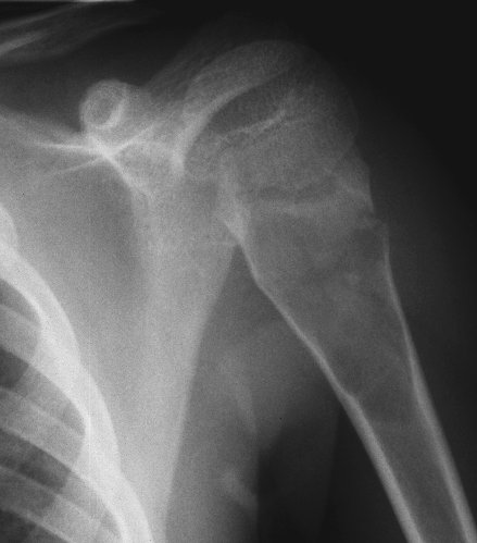
- unicameral bone cyst; simple bone cyst
- fluid-filled cavity
- fallen fragment sign: detached fragment of bone in dependent portion of cyst
- males:females 2:1
- calcaneus, long bones (esp children)
- lucent, well-demarcated, geographic
- long axis parallel to long bone
- broader toward metaphysis than diaphysis
- pathological fractures
Bosniak CT Classification of Cystic Masses
I = simple cyst; nonoperative
II = septated, minimal calcium, nonenhancing high-density cysts, infected cysts; nonoperative
III = multiloculated, hemorrhagic, dense calcification, non-enhancing solid component; renal-sparing component
IV = marginal irregularity, enhancing solid component; Radical nephrectomy
Thin Walled Cysts in the chest
- Eosinophilic granuloma
- Lymphangioleiomyomatosis
- Tuberous sclerosis
- Neurofibromatosis
Cystic Pancreatic Masses
- Pseudocyst (MC)
- True Cyst
- Lymphoepithelial Cyst
- Peripancreatic cyst
- Serous Cystadenoma
- Solid tumor with cystic change
- Mucinous Cystic Neoplasm
- Intraductal Papillary Mucinous Neoplasm
- Papillary and Solid Epithelial Neoplasm (pseudopapillary tumor of the pancreas)
Cystic Liver Masses
- Simple cyst
- Polycystic Liver
- Bile Duct hamartoma
- Caroli Disease
- Sarcoma, undifferentiated embryonal
- Biliary cystadenoma
- Neoplasm: cystadenocarcinoma, biliary cystadenoma
- Mets, cystic
- Abscess
- Hydadit cyst
- Extrapancreatitic pseudocyst (pancreatitis)
- Hematoma
- Biloma
Cystic Kidney Masses
- Polycystic kidney disease
- Simple Renal cysts
- Von Hippel Lindau disease
- tuberous sclerosis
- Uremic Cystic Kidney Disease (acquired vs medullary)
- Medullary Sponge Kidney
- Multicystic dysplastic Kidney
- Infection: TB, echinococcus, abscess
- Cystic degeneration of renal tumor


