Archive for June 2008
Don’t Touch Lesions
Postraumatic
- myositis ossificans
- avulsion injury
- cortical desmoid
- degenerative geodes
- discoid vertebral sclerosis
- healing fracture callus
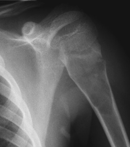
- pseudodislocation of the humerous (fracture and hemarthrosis)
Normal Variants
- dorsal (articular) lucent defect of patella
- pseudocyst of humerus
- os odontoideum
Benign Lesions
- Nonossifying fibroma
- bone islands
- unicameral bone cyst
- bone infarction


Barton’s fracture
 intra-articular radial fracture
- dorsal angulation
- plus radiocarpal dislocation
- see Colles’ fracture

Smith fracture
 fracture of distal radius and ulna with volar angulation
- opposite of Colles’ fracture

Colles’ fracture

- fall on outstretched hand
- fracture of distal radius and ulna with dorsal angulation
- metaphyseal, epiphyseal region
- see Frykman classification
- opposite of Smith fracture
- see Barton’s fracture (+dislocation)

Fracture of scaphoid

- High potential for AVN to the proximal fragement (blood supply begins distally)
- Trauma with pain over the snuff box
- If high clinical suspicion and negative film, cast and refilm in 1 week:
- Diffuse osteopenia and hyperemia around the fracture site
- Or just get MRI
- AVN = increased density of proximal pole of scaphoid
Rotary subluxation of the scaphoid
 rupture of scapholunate ligaments causing scaphoid to rotate dorsally
- AP view = Terry Thomas sign (David Letterman sign)


Hook of the Hamate fracture

- Fall on outstretched hand, athletes who hold bats or rackets that lever off hook
- Very rare, requires carpal tunnel view to see
- Bony protruberance off the hamate on the ulnar aspect of the carpal tunnel
- Do CT if strong clinical suspicion and plain film negative
Lunate and Perilunate dislocation
 best seen on lateral wrist view
- fall on outstretched hand


Frykman classification
describing Colles’ radial and ulnar fractures
Type 1 = R, extra-articular
Type 2 = R + U, extra-articular
Type 3 = R, intra-articular radiocarpal
Type 4 = R + U, intra-articular radiocarpal
Type 5 = R, intra-articular distal radioulnar joint
Type 6 = R + U, intra-articular distal radioulnar joint
Type 7 = R, intra-articular both radiocarpal and radioulnar joints
Type 8 = = R + U, intra-articular both radiocarpal and radioulnar joints
Medial Collateral Ligament
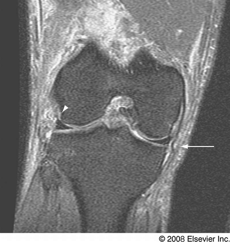
“wavy” appearance on MR
prevents valgus angulation
2 layers separated by small bursa:
- tibiocollateral ligament (superficial)
- meniscofemoral and meniscotibial ligaments (deep)
grade 1
- surrounding edema
grade 2
- thickening
- partial rupture
grade 3
- complete rupture
- loss of ligament continuity
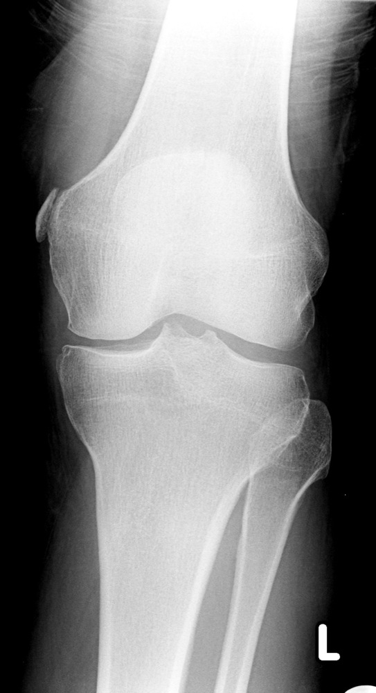
Pellegrini-Stieda = post-traumatic calcification around MCL origin related to prior trauma
Brodie’s Abscess
 infection becomes chronic
- radiolucent, well-defined geographic lesion with surrounding diffuse sclerosis
- 2/3 in metaphysis, 1/3 in diaphysis

AC joint separation
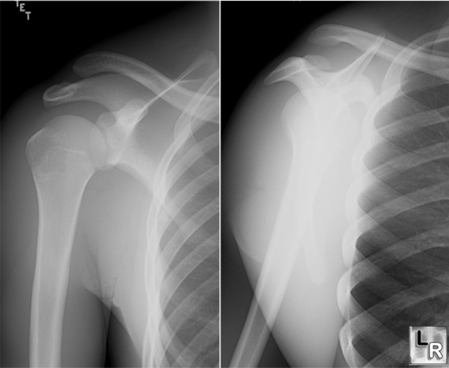
- complicaitons
- hepterotopic calcifaction-ossification
- posttraumatic osteolysis of distal clavical
- secondary osteoarthritis
- get weight bearing views, and comparison with other shoulder
Types
I = Sprain
- stretching of the AC ligament
- AC joint is stable
- CC ligament intact
- only seen on stress views, widening of AC joint
II = Subluxation
- partial or complete rupture of AC ligament
- partial disruption of CC ligament
- widened AC (less than 5 mm or 50%), normal CC
- arthroplasty may be needed
III
- disruption of both AC and CC (more than 5 mm or 50%)
- widening of AC and CC on routine film
IV = posterior
- AC and CC ligaments disrupted
- Coracoacromial ligament intact
- distal end of clavical inferior and posterior to acromion (axillary views)
V = superior
- Type IV plus sternoclavicular separation
- marked widening of AC and CC, sternoclavicular dislocation
VI = inferior
- distal end of clavicle displaced inferiorly and lodges in biceps and coracobrachialis muscles
- distal end of clavicle lies inferior to acromion
Simple Bone Cyst
- unicameral bone cyst; simple bone cyst
- fluid-filled cavity
- fallen fragment sign: detached fragment of bone in dependent portion of cyst
- males:females 2:1
- calcaneus, long bones (esp children)
- lucent, well-demarcated, geographic
- long axis parallel to long bone
- broader toward metaphysis than diaphysis
- pathological fractures


